AIF Seeks Applications for Talented Electron Microscopist

The Analytical Instrumentation Facility (AIF) seeks a talented and industrious experimentalist to join our team as an Electron Microscopy Specialist. The AIF is NC State’s primary shared facility for materials characterization with a mission to enable and lead state-of-the-art research through acquisition, development, maintenance, training, and access to major analytical and materials characterization instrumentation. Through the support of engaged faculty and experienced staff, the AIF supports state-of-the-art scanning and transmission electron microscopes, X-ray scattering and spectroscopy instruments, mass spectrometry, scanning probe microscopy, nanoindentation, and extensive sample preparation facilities. The AIF is a core nanotechnology user facility in the new Research Triangle Nanotechnology Network (RTNN), a site in the National Nanotechnology Coordinated Infrastructure (NNCI).

Primary responsibilities of the new position include training new users (both internal users from NC State and those external to NC State) as well as performing service work for external clients. The ideal candidate will be customer-focused and exhibit a commitment to excellence in all technical and organizational aspects of their role. The new Postdoc will work closely with the AIF and RTNN teams in serving the needs of university, industrial, and government researchers from across NC State, the North Carolina Research Triangle, and the nation.

Please encourage talented applicants to apply:
Full-time staff position: https://jobs.ncsu.edu/postings/76529
Postdoc: https://jobs.ncsu.edu/postings/76522

Image Contest Winner – Yaewon Park

Congratulations to our image contest winner, Yaewon Park, for her entry, CaCO3 mineralized poly(vinyl alcohol) nanofibers.

This picture shows a Scanning Electron Microscopy (SEM) image of CaCO3 nanoparticle clusters encrusting electrospun poly(vinyl alcohol) nanofibers. This structure resembles bone structure which consists of collagen fibrils and hydroxyapatate crystals attached along them.

My current research is on surface coating of nanofibers with CaCO3 particles by mimicking bone formation process. My research is expected to give a light on environmentally friendly coating of functional textiles and water filtration materials. Nanofibers were dipped in CaCl2 solution and Na2CO3 solution alternatively for 10 times. This image shows that spherical CaCO3 particles surrounded the circumference of nanofibers. This interesting structure is similar to human bone structure.

Honorable Mention Images:
img-2-coral-reef
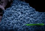
Joshua Zhou: Coral Reef The viewing window of a scanning electron microscope halts before a field of “coral reef”, ordered clusters of vanadium oxide nanorods. Another rod rests on their surface, like a fish seeking shelter from predators. Characterizing the shape of vanadium oxide nanomaterials can account for changes in their thermochromic properties.

This work aimed to form a titanium oxide-vanadium oxide composite doped with magnesium in order to increase the infrared blocking capability of thermochromic films. Efficient thermochromic films can be used in smart windows to block heat-bearing infrared radiation on hot days, while phase-shifting in cold weather to allow warmth in from sunlight. Phase shifts are temperature dependent and rely on no external supply of electricity. This can help reduce air-conditioning bills while maintaining room comfort.

mn3
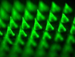
Yanqi Ye: Smart Melanoma Patch Fluorescence imaging of a representative microneedle patch that contained FITC-aPD1 loaded NPs for melanoma treatment. Despite recent advances in melanoma treatment through the use of anti-PD- 1 (aPD1) immunotherapy, the efficacy of this method remains to be improved. Here we report an innovative self-degradable microneedle (MN) patch for the sustained delivery of aPD1 in a physiologically controllable manner. Moreover, this administration strategy can integrate with other immunomodulators (such as anti-CTLA- 4) to achieve combination therapy for enhancing anti-tumor efficacy.


Nano Fabric Breaks Down Chemical Warfare Agents

Researchers from the lab of RTNN principal faculty member Greg Parsons have created a material capable of degrading chemical warfare agents (CWAs). Uniform coatings of metal-organic frameworks (MOFs) were grown on electrospun nanofibers, forming unique kebab-like structures. These MOFs were able to break down CWAs making them harmless. The team conducted much of their characterization work at the Analytical Instrumentation Facility, a member of the RTNN. More information about the work can be found below and in the NC State Press release.

“Ultra-Fast Degradation of Chemical Warfare Agents Using MOF–Nanofiber Kebabs”
Junjie Zhao, Dennis T. Lee, Robert W. Yaga, Morgan G. Hall, Heather F. Barton, Ian R. Woodward, Christopher J. Oldham, Howard J. Walls, Gregory W. Peterson, and Gregory N. Parsons.

Abstract: The threat associated with chemical warfare agents (CWAs) motivates the wardevelopment of new materials to provide enhanced protection with a reduced burden. Metal–organic frame-works (MOFs) have recently been shown as highly effective catalysts for detoxifying CWAs, but challenges still remain for integrating MOFs into functional filter media and/or protective garments. Herein, we report a series of MOF–nanofiber kebab structures for fast degradation of CWAs. We found TiO2 coatings deposited via atomic layer deposition (ALD) onto polyamide-6 nanofibers enable the formation of conformal Zr-based MOF thin films including UiO-66, UiO-66-NH2, and UiO-67. Cross-sectional TEM images show that these MOF crystals nucleate and grow directly on and around the nanofibers, with strong attachment to the substrates. These MOF-functionalized nanofibers exhibit excellent reactivity for detoxifying CWAs. The half-lives of a CWA simulant compound and nerve agent soman (GD) are as short as 7.3 min and 2.3 min, respectively. These results therefore provide the earliest report of MOF–nanofiber textile composites capable of ultra-fast degradation of CWAs.

2016 Nobel Prize in Chemistry Awarded for Nano-Machines

The Nobel Prize in Chemistry was awarded to three scientists for their work in the creation of “nano-machines.” Jean-Pierre Sauvage of the University of Strasbourg, Sir Fraser Stoddart of Northwestern University, and Bernard Feringa of the University of Groningen. Sauvage demonstrated the ability to flexibly link molecules, Stoddart discovered how to make the molecules move, and Feringa determined how to power them. The combination of these discoveries resulted in molecular machines, nano in size, whose movements are controllable. These nano-machines can be used to perform specific tasks and are being explored for use in numerous fields including medicine, computing, and energy.

For more information about their exciting discoveries please visit the Nobel website.

RTNN acquires portable Scanning Electron Microscope

Through collaboration with the Parsons Research Group at NC State, the RTNN now has available a PhenomWorld Desktop SEM. The Phenom SEM allows for very fast microscopic imaging of samples in a compact, portable package – about the size of a desktop computer. The RTNN will use the Desktop SEM in outreach programs, such as bringing instruments to classrooms for demonstrations of instruments used in nanoscience. If you are interested in having the RTNN bring the desktop SEM to your event or school, please contact Phillip Strader (phillip_strader@ncsu.edu) or Maude Cuchiara (maude_cuchiara@ncsu.edu)